Page Contents
OVERVIEW
How could one every complicate the diagnosis of exertional rhabdomyolysis? This topic in medicine seems fairly straightforward. In settings of extreme exertion (such as strenuous exercise) a large amount of muscle cells are damaged, releasing their contents into the patient’s serum. This can cause the patient to experience various symtpoms (muscle swelling, nausea, malaise), can cause changes to the color of the patient’s urine (“tea” colored urine can be caused by myoglobinuria), and lab values can also suggest skeletal muscle damage (most notably creatine kinase levels are elevated in the patient’s serum).

While there are a few components to exertional rhabdomyolysis…it really does not seem to be too complicated. So this begs the question, how could the diagnosis of such a simple condition ever become complicated? Read on to answer that question with a real example.
WHAT HAPPENED?
The patient in question is a 30 year old woman who does not have any remarkable past medical history. She is admitted to the hospital with a creatine kinase (CK) value that is off the charts (> 40,000). It was known that about 2 weeks ago the patient went to the ER because she had noticed unusual swelling in her muscles after a strenuous arm workout. At the time her CK value was elevated (~7,000) however other then her swollen arms she was asymptomatic and sent home with the diagnosis of exertional rhabdomyolysis. The patient was told not to exercise excessively for the time being, and was instructed to follow up in 1 week with her primary care physician (PCP) so her CK value could be checked again (to ensure that it had gone down).

A week later, the patient returns for her follow up appointment and her CK value is checked. It is discovered that her CK value has risen to >40,000. Given that the patient is presumed to have not exercised in the past week, it is thought that this patient might have a rheumatological or metabolic cause of her elevated CK value, and the diagnosis of exertional rhabdomyolysis is quickly abandoned. She is told that she does not need to come into the ER, but will require further workup with the help of a rheumatologist given her “exertion-indepenednet” rise in CK.
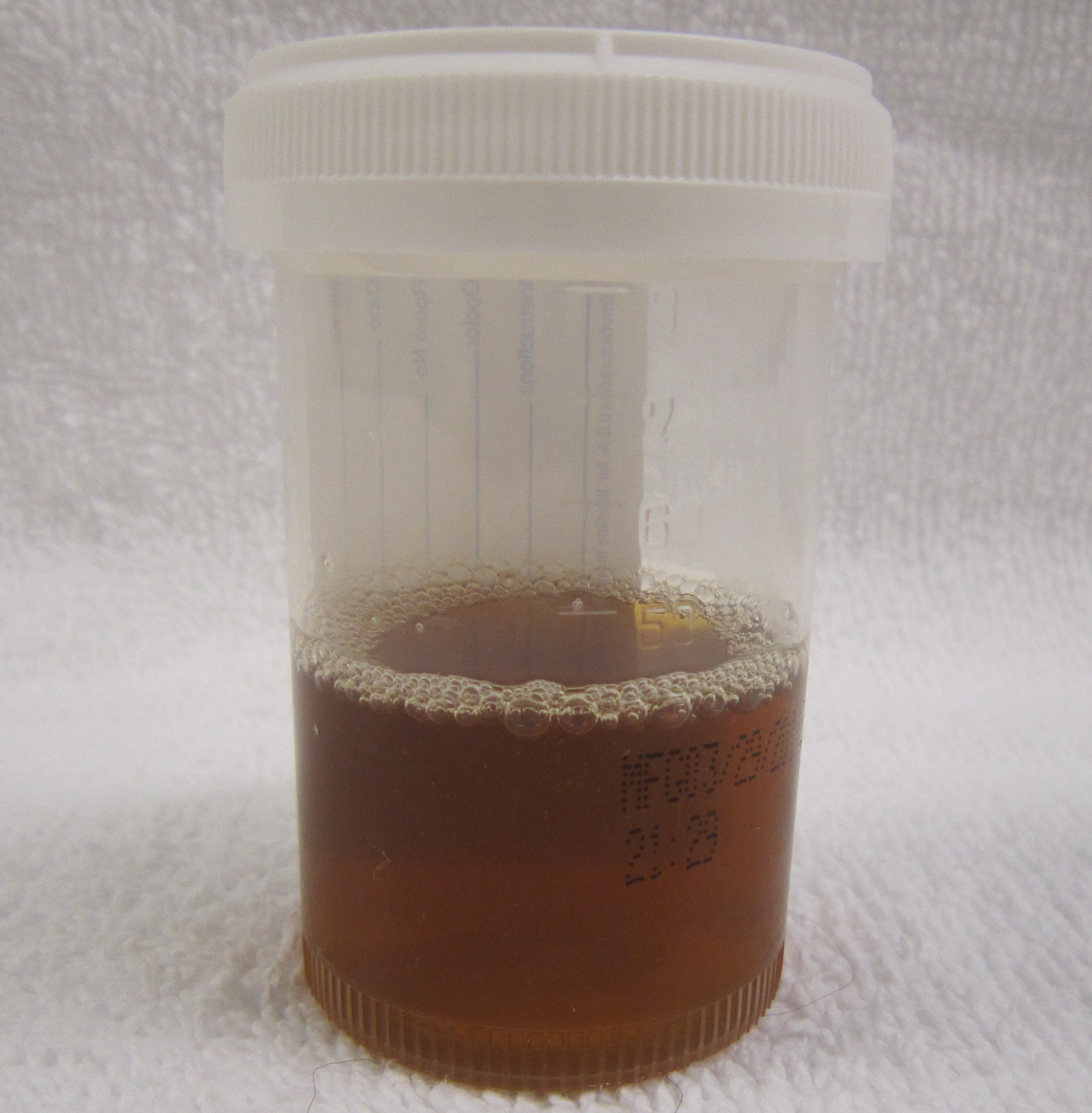
Later that night, the patient begins to feel odd symptoms. Her legs begin to swell noticeably, she gets dizzy, and feels the general sense of malaise. Upon passing urine she notices that it is now red colored and decides to go back to the ER. Here, given her past history, symptoms, and sharp rise in CK, the patient is quickly admitted to the medicine floor for workup and management.

Upon admission, an extensive rheumatological workup process is begun. Diseases such as polymyositis and even rare conditions such as mutations in the RYR1 gene (that predispose to muscle damage/malignant hyperthermia) are seriously considered. Plans are put into place to try and sequence the patient for mutations, and an EMG informed muscle biopsy is even suggested if the patient’s creatine kinase values remain elevated. The patient becomes concerned that she might have a chronic illness and does not understand how this is possible, given that she has never experienced any type of symptoms before, and has no family history suggestive of genetic conditions that run in her family.
At this point during the workup, it is revealed that the patient actually had never fully refrained from exercising during the period of time in between her ER visits. It becomes quite clear that her jump in CK (from 7,000 to >40,000) can actually be explained by a very intense lower body workout (which now explains why the patient noticed swelling in her legs prior to coming to the ER a second time. With this new information it seems very obvious that this presentation is still actually very consistent with exertional rhabdomyolysis, and that the rheumatological workup for the patient’s elevated CK was out of turn.
The patient is diagnosed with exertional rhabdomyolysis, and is monitored and is in the hospital. Now that she is on the floor she is no longer exercising. Her CK numbers become lowered, and she is discharged from the hospital when the value is <20,000. There is no need for any further workup (muscle biopsy, genetic sequencing) at this time, and the patient is educated on pacing herself during exercise to avoid future issues with excessive muscle damage.
AT WHAT POINT DID THE FOREST BECOME LOST IN THE TREES?
It seems that at the follow up appointment things seem to shift into a different light. Because the PCP of the patient quickly assumed that she had stopped exercising, the workup for the patient quickly shifted to uncovering the “non-exertional” cause of the patients elevated CK. This is not advantageous simply because the cause of the patient’s rhabdomyolysis was in fact exertional! This very subtle change in thought process completely changes the clinical workup, as a more extensive means of investigation is needed to understand what could explain the patient’s clinical picture.

WHY SHOULD WE NOT THINK ITS COMPLETELY “CRAZY” THAT THIS HAPPENED?
While it can be easy to blame the PCP for mistakenly “mis-framing” the patient’s history, it is important to take a step back and realize how it might be possible to make such a mistake.
Given that the patient was instructed not to exercise intensely after her initial rise in CK, we can appreciate that the physician would not expect for the patient to have engaged in such an intense workup prior to her stark rise in CK. What’s more, the patient’s rise in CK seems to be so dramatic (it is effectively over a 4x increase in serum levels), that it seems as though it might be caused by something other then exertion alone.
Fundamentally there was a miscommunication between the physicians and the patient in understanding the importance of her recent exercise. One may also feel inclined to blame to the patient for “withholding” relevant information, however upon conducting the interview it was clear that the patient did not realize how critical this piece of information was for the clinical workup. Sometimes things move so fast in medicine that we barely have time to think clearly as to what is going on.
WHO CARES? WHAT WAS THE HARM IN WHAT HAPPENED?
While this patient was ultimately diagnosed correctly, let us not underestimate the opportunity cost of pursuing the rheumatological workup for this patient’s elevated CK values:
- Compromised patient care: the patient’s care becomes influenced by the suspected etiology behind her elevated CK values.
- Extended hospital stay: the more time a patient spends (unnecessarily) in the hospital the higher their rate of infection/development of other complications. The confusion around this patient’s CK elevation led to a long hospital stay.
- Expenses: all the studies ordered have a financial cost associated with them. Whether it is the hospital, insurance companies, or even the patient, someone has to foot the bill of ordering genetic testing, biopsies, and rheumatological studies. Luckily many of these tests were never ordered to pursue diagnosing conditions such as polymyositis/metabolic causes of CK elevation.
- Time: everyone involved in the care of the patient (and the patient themselves!) will lose time the longer a workup drags on (and if unnecessary tests are ordered). This time could be put to better use in caring for other patients, and there is always a premium on time in medicine!
WHAT IS THE TEACHING POINT HERE? HOW DO WE AVOID THIS IN THE FUTURE?
This is yet another clear example of how a good patient history can really help with the management of a patient. In the case of this patient’s presentation, upon her stark elevation in CK it is very important for the physician interviewing the patient to clarify exactly what role exercise/exertion could have had in the development of her symptoms/the results of her lab work.

Had the medical team known that this patient had only experienced this CK increase AFTER a very strenuous lower body workout (a few days prior to her admission) her workup would have been much more brief. Considering rheumatological/metabolic causes would have not been necessary, and much time/effort could have been saved by everyone.
It is important to realize that their is always the slight chance that this patient may have a chronic condition that predisposes her to muscle damage (such as a mutation, inflammatory condition) however given her pattern of symptoms only being associated with extreme exercise this is very unlikely and does not seem worth considering. If in the future the patient develops a similar presentation, truly independent from strenuous exertion, then these causes can be pursued more fully.
Page Updated: 09.27.2016