Page Contents
WHEN DO WE DO IT?
The abdominal exam can be performed during a regular screening, and can also be performed during a focused exam. Patient presentations that might further indicate the need for an abdominal exam can include:
- Pain in the abdomen
- Vomiting
- Diarrhea
- Constipation
*This list is by no means exhaustive, if time allows try to perform as many components of the physical exam as you can
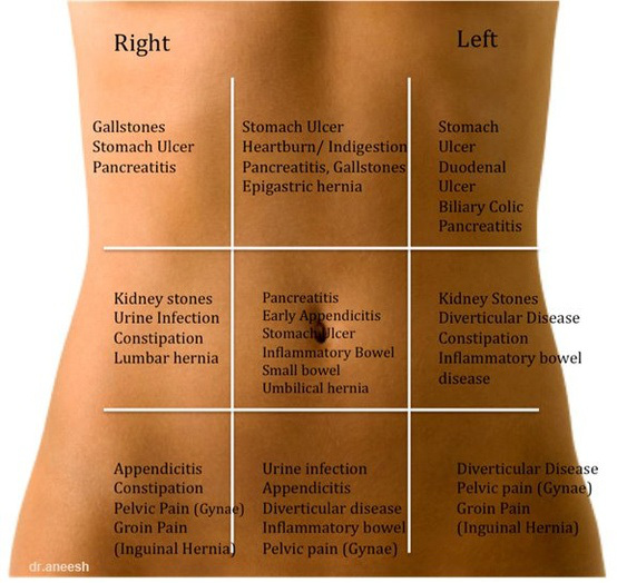
COMPONENTS
The abdominal has a few key components that we will discuss in greater detail below (that should be completed in the following order):
- Inspection
- Auscultation
- Percussion
- Palpation
- Supplemental exams (as indicated)
INSPECTION
The exam beings with its least invasive component: visual inspection. While at this point you likely will have taken a thorough past medical history, you can use this as an opportunity to ask more questions that relate to the abdomen. Take note of the following things:
Overall appearance: is the abdomen symmetrical? Is the abdomen swollen or distended?
Scars: Does the patient have any scars (either from trauma/surgery)? Remember each scar has a story behind it and can be worth asking about.
Dermatological findings: are there any suspicious rashes, moles, skin findings? While the patient has their abdomen exposed you might as well take advantage of conducting a portion of your skin exam as well.
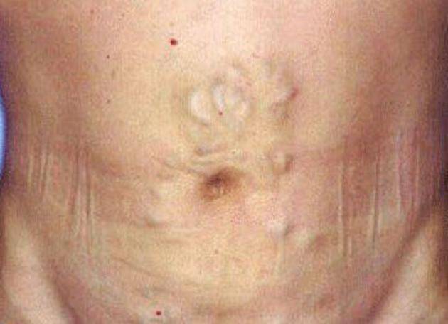
Other abnormalities? these can include a wide variety of findings such as: hernias, swollen vessels (caput medusae), wounds, infections, bruises etc.
AUSCULTATION
After visually inspecting, using our stethoscope we then listen to the abdomen.
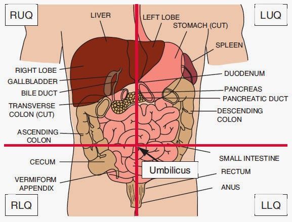
Bowel sounds: typically listening to bowel sounds is the most useful when trying to investigate the suspicion of a bowel obstruction (absent or high pitched bowel sounds can be observed). Listening to all 4 quadrants of the abdomen can be beneficial in order to ensure that bowel sounds are present throughout.
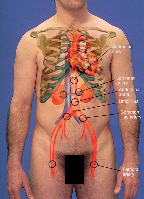
Bruits: refers to a vascular murmur that is caused by turbulent flow. Given that you already have your stethoscope out and touching the patient’s abdomen you can quickly check for the presence of bruits as well (various arteries). Here is an example of a renal bruit.
PERCUSSION
Percussion relies upon you to use your hands as literally a percussive instrument (and discern the difference in sound as a means for appreciating underlying structures). Hollow sounds will suggest the presence of air, while more dense sounds can be suggestive of organs underneath the site of percussion.
Technique: the technical aspect of percussion is one that eludes many individuals. While silly, the following video does explain the technical aspects of percussion well. A few things to keep in mind.
- Keep your nails short (you want the pad of your finger to make contact when percussing, not your nail)
- Keep the finger you are touching to the patient flat and flush against their skin.
- Flick your wrist quickly (this is where the force should be generated from
Tympany: if time allows, the whole abdominal surface can be percussed to assess for pockets of air. While doing this, keep an ear out for any unusual dullness as well that might be suggestive of fluid in the peritoneum (such as ascites).
Liver Percussion: one of the organs we can effectively percuss in order to determine its size. Percussing the liver along the midclavicular line is usually sufficient. Areas of percussion that reside above the liver have a duller sound.
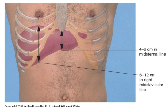
*One can measure on their hand how long 12 cm is and use this as a crude means of assessing the liver size.
Spleen percussion: if the spleen significantly enlarged, it can alter percussion (increased dullness) in the field above its location (left upper quadrant).
PALPATION
This is the most “hands on” portion of the exam, and you can begin with one finger (especially if you have a patient who is in pain).
Deep palpation: palpate the entire abdominal surface (using both hands) if possible and take note of the following things:
- If any pain is experienced by the patient
- If the patient guards (tenses the abdomen)
- Tenderness
- Rigidness that is not due to guarding
- Masses
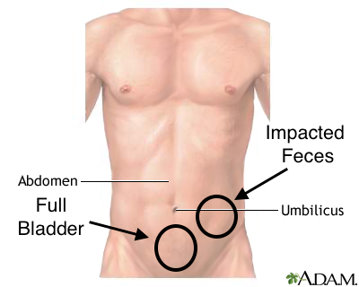
Liver palpation: upon inspiration either the tips of all the fingers OR the side of the index finger can be pushed under the rib cage to feel for the liver. Feeling the liver with this maneuver might suggest an enlarged liver. Here is a video demonstrating the maneuver.
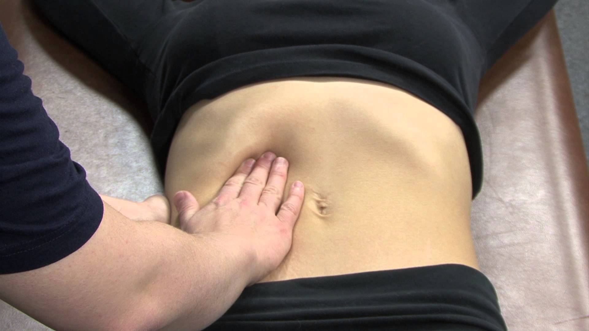
Spleen palpation: the spleen in palpated in a similar fashion, however the left hand is used to pull the rib cage towards the examiner, as the right hand is pushed under the ribs to palpate for splenomegaly (during inspiration). The spleen is placated initially in the supine position.
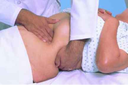
However in addition the spleen is also palpated in the decubitus position (with the patient rolled on their side).
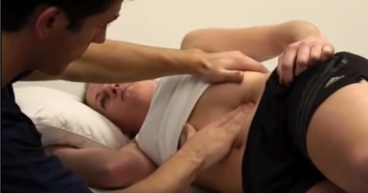
Rebound tenderness: this maneuver is conducted by applying pressure to an area, then immediately releasing the pressure. If more pain is elicited with the release of pressure (then its initial application) the test is said to be positive. This is suggestive of peritonitis, and can also be positive in appendicitis.
SUPPLEMENTAL TESTING
The below are additional tests that can be conducted if there is a clinical suspicion of specific disease pathologies.
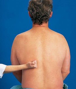
Flank tenderness: percussion can be conducted on the back of the patient above the kidney (lightly beating the bottom of the fist against the flank) to assess for the presence of kidney pain.
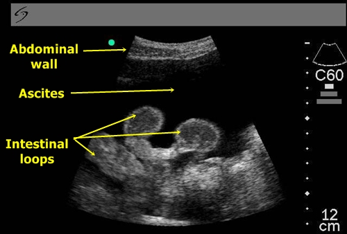
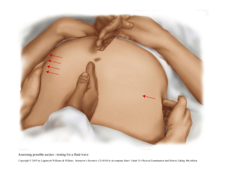
Ascites: while ultimately an ultrasound can help visualize ascites very accurately and easily, there are physical exam maneuvers that can be conducted to heighten our clinical suspicion for it.
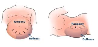
Shifting dullness test: if there is ascites in the patient, there will be increased dullness upon percussion that will “shift” as the patient is moved side to side (because the fluid will move relative to gravity). This type of shifting dullness will suggest the presence of ascites.
Fluid wave test: have the patient (or an assitant) fix the midline of their abdomen in place (using the side of their hand, they should apply pressure to their abdominal midline in order to keep it in place). Using one place your hands on either side of the abdomen (on the obliques of the patient). Tap one side while feeling for a fluid wave on the other. If a pressure wave is transmitted this is suggestive of fluid within the peritoneum. Demonstration here.
Page Updated 01.17.2016