Page Contents
OVERVIEW
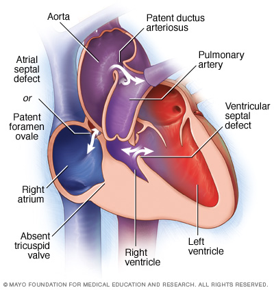
Tricuspid Valve Atresia (TVA) refers to a congenital heart condition that is characterized by the complete lack of a tricuspid valve. This lack of communication between the right heart chambers results in a hypo plastic right ventricle.

Other associated features include:
- Patent foramen ovale/atrial septal defect
- Ventricular septal defect
- Pulmonary stenosis (from decreased blood flow out of the pulmonary valve)
WHY IS THIS A PROBLEM?
Because the tricuspid valve is absent NO BLOOD WILL TRAVEL DIRECTLY FROM THE RIGHT ATRIUM TO THE RIGHT VENTRICLE. As a result, less blood will be pumped out to the lungs.
WHAT MAKES US SUSPECT IT?
Initial Presentation
- Cyanosis in newborns
Physical Exam:
Cardiac exam:
- Holosystolic murmur can be caused by ventral/septal defect
CLINICAL WORKUP
Chest X-Ray: while not a diagnostic study, a chest radiograph can reveal the following features of TVA:
- Decreased pulmonary markings: this is due to the lack of blood flow to the lungs.
Electrocardiogram: the cardiac changes seen in TVA can be reflected on a EKG study (in newborns):
- Left axis deviation: typically newborn should have right axis deviation given that blood is shunted away from the left side of the fetal heart (and the right side is larger in newborns)
- Small/absent R waves in precordial leads: similar reasons as to why left axis deviation is seen in TVA.
- Tall/peaked P waves: the atrial spatial defect permits increased blood flow to the right atrium which causes its enlargement (in turn causing this EKG finding).
HOW DO WE TREAT IT?
Surgical correction:
ARCHIVE OF STANDARDIZED EXAM QUESTIONS
This archive compiles standardized exam questions that relate to this topic.
Page Updated: 11.23.2016