Page Contents
OVERVIEW
This page is dedicated to organizing various examples of standardized exam questions whose topic is glioblastoma multiforme (GBM). While this may seem a odd practice, it is useful to see multiple examples of how GBM will be characterized on standardized exams (namely the boards and the shelf exams). This page is not meant to be used as a traditional question bank (as all of the answers will be the same), however seeing the classic “test” characterization for a topic is quite valuable.
KEY CHARACTERISTICS OF THIS CONDITION (ON EXAMS)
When it comes to standardized exams, each topic has its own “code” marked by key buzzwords, lab findings, clues, etc. If you are well versed in this code you will be able to more quickly identify the condition that is being discussed, and get the right answer on the exam you are taking. Below is the “code” for GBM.
Chief Complaints:
- Headaches
- Nausea
- Seizures
Clinical Workup:
- Often crosses hemispheres through corpus callosum: “butterfly” lesion
- Lesions enhance with contrast on imaging
- Histology:
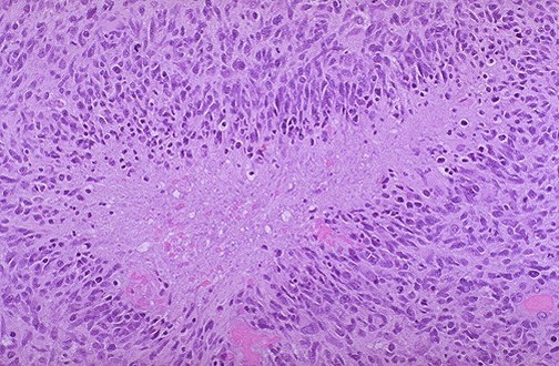
- “Pseudopalisading tumor cells” can be seen on a brain biopsy
- Necrosis and hemorrhage in the center of the tumor
- GFAP positive on immunofluorescence
QUESTION EXAMPLES
Question # 1
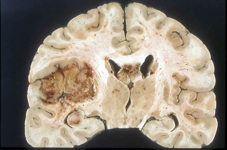
A 60 year old male comes to the ER after the new consent of generalized tonic-clonic seizures. His partner explains that for the past few weeks he has been complaining of headaches, nausea, and weakness. He has even been experiencing the headaches at night, and they have become so severe that they wake him up from his sleep. The patients past medical history is unremarkable other then a diagnosis of osteoarthritis. He is admitted to the hospital for further workup. He develops septic shock from a hospital acquired pneumonia and passes away. A brain section is performed and shown below:

What is the likely diagnosis in this patient?
Explanation # 1
Brain lesion + areas of necrosis/hemorrhage seen on gross histology = GBM
Question # 2
A 70 year old male is brought to the emergency room after he undergoes new tonic-clonic seizures. His brother explains that recently the patent underwent personality changes to the point that he is a “entirely new person”. A CT-scan of the head reveals the presence of a single brain lesion that enhances with contrast. It is found on the left hemisphere of the brain, and crosses over to the right side through the corpus callosum. What is the likely diagnosis in this patient?
Explanation # 2
Brain lesion + enhances with contrast + butterfly lesion (crosses corpus callosum) = GBM
Question # 3
A 65 year old male develops the new consent of headaches. An MRI is performed and reveals the presence of a intracranial mass that is biopsied for further analysis. Light microscopy from the lesion is shown below:

What is the likely diagnosis in this patient?
Explanation # 3
Brain lesion + pseudopalisading tumor cells on histology = GBM
TESTABLE FACTS ABOUT THIS TOPIC (BEYOND ITS IDENTIFICATION)
Many questions on standardized exams go beyond simply recognizing the underlying topic. Often there are specific testable facts regarding some aspect of the topic’s pathophysiology/management/clinical implications that are commonly asked. Some of these are listed below:
Cause:
- Tumor cell origin: astrocytes (this condition is classified as a grade IV astrocytoma)
Page Updated: 05.01.2017