OVERVIEW
Interpreting a chest X-ray is a very valuable skill given how commonly it is ordered. The following guide helps walk you through some of the basic elements of X-ray imaging that are important to understand in order to interpret these studies effectively.
HOW DOES IT WORK?
This imaging modality works by literally penetrating the human body with X-rays (and measuring X-rays that can not penetrate the medium in question). If the X-rays penetrate a structure, they will appear black on the film. If they can’t penetrate the structure (i.e. are reflected) they appear white on the film.
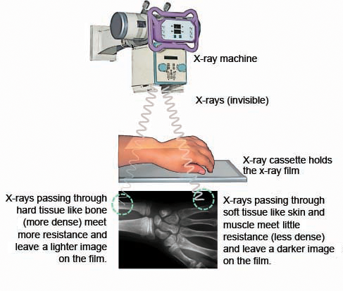
There are 4 major radiological/physical mediums that appear different visually given their relative densities. They are (listed from least to most dense):
- Air: this will appear black because the X-rays can easily penetrate air
- Fat: fat will not be completely penetrated and will appear faintly on film
- Water (soft tissues): this medium will appear more white on X-rays (compared to fat) given its increased density.
- Bone (calcium): bony/calcified structures show up the most clearly on X-rays (bright white!) because the X-rays have difficulty penetrating these structures.
TYPE OF X-RAY MACHINE
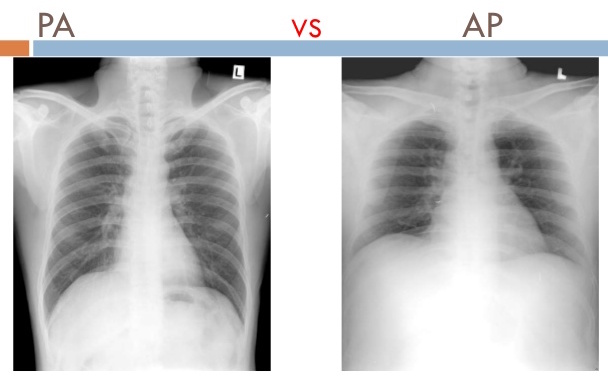
When looking at an X-ray image, arguably the first detail you should notice is if the image was taken with a portable or non-portable device. Portable devices are advantageous because they can taken to a bed ridden patient, however non-portable X-rays are preferred (when a patient is able to ambulate) because of their superior image quality. It is important to be wary of comparing portable to non-portable X-rays simply because there are differences between the two (a patient’s pneumonia may look worse on a portable X-ray compared to a baseline non-portable image). We will discuss quality measures for X-rays more below.
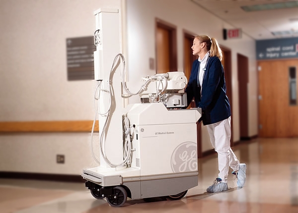
***Keeping this type of X-ray machine in mind will contextualize your analysis of the image further on.***
PENETRATION OF THE FILM
The penetration of the film refers to how well the X-rays have penetrated the body of the patient. The more penetration the more X-rays will interact with the film placed on the other side of the patient. We must appreciate that their is a “sweet spot” with regards to how well penetrated the film is (to maximize the clinical information we can gain from the study). Below are some examples from a chest X-ray study to help demonstrate this concept.
Is the film under-penetrated? If film is under penetrated then there will be an excess of white present.
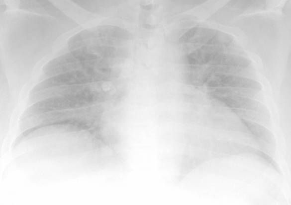
Is the film over-penetrated? Alternatively, if the X-rays over penetrate the patient, there will be an excess of black present on the film.
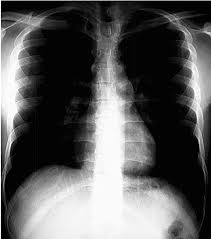
***PROPER PENETRATION IS CRITICAL TO INTERPRETING AN X-RAY!***
Magnification: How far away is the structure from the film when the image was taken? This can be a difficult concept to retain, however the further away a structure is from the X-ray film, the more it will MAGNIFY as the X-ray film is developed. It is for this reason that a standard PA (with the heart closer to the film) shows a more accurate size of the heart while an AP film (which increases the distance between the film and the heart) will show enlarged cardiac structures.
Page Updated: 08.24.2016