OVERVIEW
This page is dedicated to covering a very important call finding: cervical prevertebral edema. This page will specifically cover this diagnosis in the setting of making the finding on CT imaging. The cervical spine/neck is often times imaged as a part of trauma screening (such as a brain and cervical spine) but can also be imaged on other studies as well (such as a code stroke CT protocol which includes a CTA of the neck).
This page will focus primarily on non-contrast findings however will also encompass findings that can be apprecaited on post-contrast imaging as well.
WHAT IS IT EXACTLY?
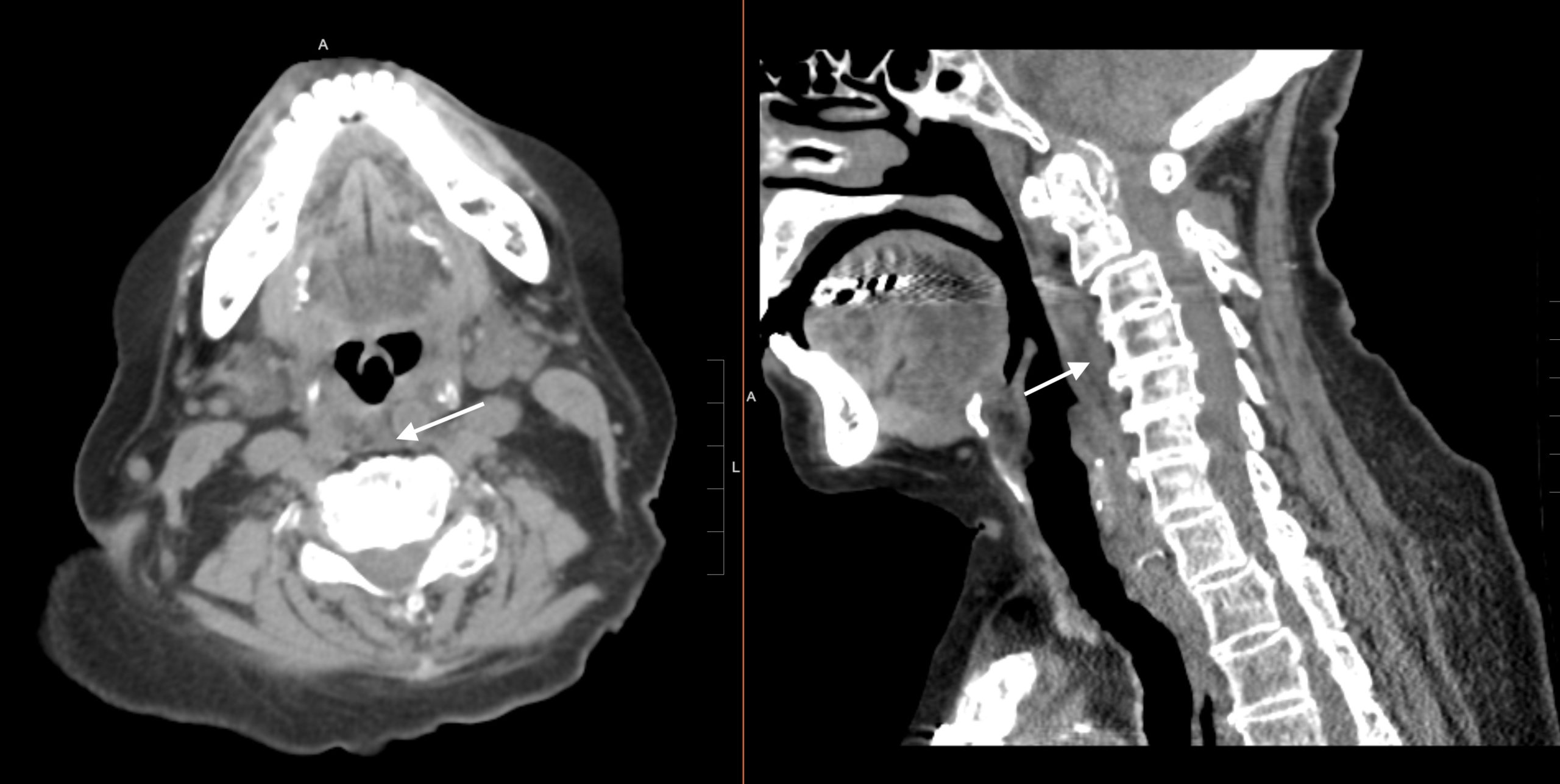
Cervical prevertebral edema is a self descriptive diagnosis. It refers to abnormal fluid/edema in the prevertebral space (often involving the fat just anterior to the vertebral bodies) of the cervical spine.
CASE ARCHIVE
Use the following login information to access the case archives: USERNAME: user, PASSWORD: password.
Click here to open up a case archive of examples of cervical prevertebral edema on CT.
Page Updated: 07.19.24