OVERVIEW
This page is designed to help cover some key radiological features of the condition Scleroderma (systemic sclerosis). Fundamentally, this condition is characterized by the deposition of collagen in areas of the body. On physical exam the hands and feet may demonstrate sclerodactyly which might warrant for imaging of these areas of the body.
Some key radiological features seen in patients with scleroderma include:
- Malposition
- Periarticular calcifications
- Articular erosions
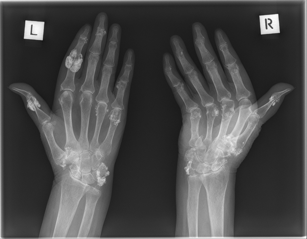
PERIARTICULAR CALCIFICATIONS IN SLERODERMA
While this radiological finding is not only specific to scleroderma, it is worth noting that often in this condition patient X-rays can demonstrate calcifications that are around joints (periarticular). Below are some examples.
Example 1:

Page Updated: 09.03.2016